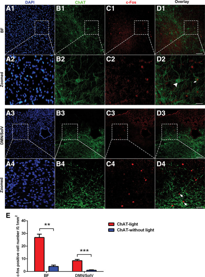
Figure 4.

Photostimulating basal forebrain (BF) cholinergic neurons induced c-Fos expression in the BF and the dorsal motor nucleus of the vagus (DMN)/ventral part of solitary nucleus neurons (SolV). A1–D1, Immunofluorescent labeling showing that photostimulating BF cholinergic neurons induced c-Fos expression in a large number of BF neurons. A2–D2, Serial images projection. Some ChAT+ neurons are c-Fos+ (arrows in D2), and some neurons are ChAT– and c-Fos+ (arrowhead in D2). A3–D3, Increased c-Fos expression was observed in the DMN/SolV. A4–D4, Serial images projection. Most neurons were ChAT–/c-Fos+ (arrowhead in D4). E, Quantification of c-Fos–positive cells in ChAT mice that were or were not photostimulated. These data are presented as the mean ± sem (**p < 0.01, ***p < 0.001; scale bar = 50 μm). DAPI = 4′,6-diamidino-2-phenylindole.
