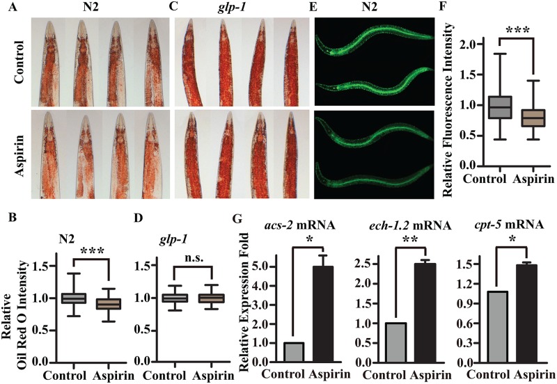
Fig 3. Aspirin increases lipid hydrolysis.
Oil Red O Staining of (A) wild-type worms and (C) glp-1(e2141)III. mutants treated with 100 μM aspirin. The relative Oil Red O intensities of (B) wild-type worms and (D) glp-1(e2141)III. mutants were calculated using Image J. (E) Aspirin decreased the absorption of BODIPY- labelled fatty acids in wild-type N2 worms. (F) The relative GFP fluorescence intensity was calculated using Image J. For box-and-whisker plots, the whiskers show the minima and the maxima within a 1.5 IQR. (G) The mRNA level of the expression of enzymes in fatty acid β-oxidation of wild-type worms treated with or without 100 μM aspirin. The figures show the mean value of three independent experiments, and the error bars represent the SEM. P values were calculated by a two-tailed t-test, * p < 0.05, ** p < 0.01. Not significant was abbreviated as n.s. Statistical details and repeats of these experiments are summarized in S4, S5 and S8 Tables.

