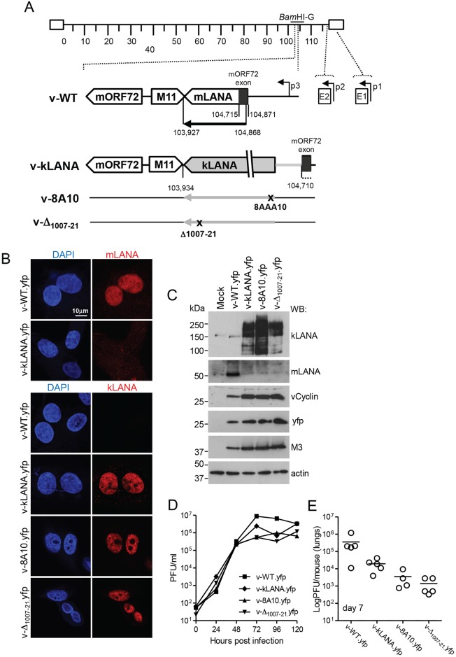
Fig 3. Generation and lytic growth of MHV68 chimeric viruses.
(A) Schematic diagram. The kLANA cassette was inserted between the M11 stop codon and the mORF72 exon in place of MHV68 103,935–104,709, which includes most of the mLANA ORF. p1, p2, p3, are mLANA promoters[38,39]. The mORF72 noncoding exon (black) is located within the mLANA coding region. The E2 splice acceptor site (nt 104,871) and the mORF72 exon splice donor site (nt 104,715) were left intact to ensure expression of kLANA and mORF72. The mLANA start codon and three downstream ATGs were mutated to ATT to prevent initiation of translation (indicated by black dots). The BamHI-G fragment (genomic nt 101,653–106,902) is indicated. mLANA ORF, nt 104,868–103,927. (B) Confocal immunofluorescence detection of mLANA (top panels) or kLANA (lower panels) from yfp viruses. Magnification 630x. (C) Immunoblot of viral proteins. (D) Growth curves of virus in BHK-21 cells after infection with 0.01 PFU/cell. There was no significant difference between infection groups (p>0.05 using one-way non-parametric ANOVA Kruskal-Wallis). (E) Lung virus titers 7 days after infection with 104 PFU of the indicated viruses. Circles represent titers of individual mice (n = 19). Bars indicate the mean. v-Δ1007-21.yfp had significantly lower titers than v-WT.yfp (**p<0.01, using one-way non-parametric ANOVA Kruskal-Wallis followed by Dunn´s multiple comparison test). There were no other statistically significant differences between groups.

