
Fig. 2.
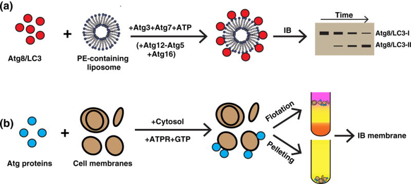
A summary of reconstituting the autophagic protein–membrane interaction. (a) In vitro lipidation. Purified lipidation components including Atg8/LC3 (C-terminal glycine exposed), Atg3, and Atg7 (plus Atg12−Atg5 and Atg16 at times) were incubated with PE-containing liposomes and ATP. The reaction led to the covalent conjugation of Atg8/LC3 to PE (and PS) on the liposome. The presence of lipidated Atg8/LC3 (Atg8/LC3 II, the higher mobility band) was determined by immunoblot (IB) (46–49,73). (b) Cell-free membrane targeting of ATG proteins. Purified ATG proteins (such as ATG14) were incubated with cytosol and cellular membrane (52,53). Alternatively, cytosol expressing ATG proteins (such as ATG14) was incubated with the cell membrane, GTP, and an ATP regeneration system (ATGPR) (54). After the reaction, the membrane fraction was collected by either centrifugation to obtain the membrane pellet or by density gradient to obtain the membrane layer. The membrane-associated ATG proteins were determined by IB.