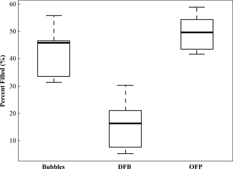
Figure 9.
Calculated percentage of kidney pixels exceeding the agent-free baseline threshold value for the different contrast agents at the 1 minute time point. Contrast generated by the activation of OFP filled a greater percentage of the kidney than a microbubble injection, while activation of DFB droplets produced an irregular contrast generation pattern and filled less of the kidney.

