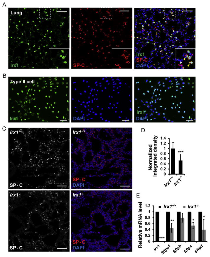
Fig. 5. Irx1 marks alveolar type II epithelial cells and regulates surfactant proteins expression.
(A) Immunostaining of Irx1 and SP-C in adult lung. Yellow arrows show Irx1+ cells are co-stained with SP-C. The white arrow shows only SP-C expression. (B) Immunostaining of Irx1 in AEC2 cell line from rats. All cells are stained with Irx1 antibody and 4′-6-diamidino-2-phenylindole (DAPI). (C) Bar chart shows the fluorescent density of SP-C in Irx1+/+ and Irx1−/− left lungs at P0 stage. p = 0.00012. (D) Bar chart shows the percentage of SP-C+ cells in Irx1+/+ and Irx1−/− left lungs at P0 stage. (E) Quantitative RT-PCR analysis of Irx1 and surfactant proteins mRNA level in the left lung of E18.5 stage embryos. n = 6, *, p < 0.05; **, 0.01 < p < 0.05; ***, p < 0.001. Scale bar: 50 μm. (For interpretation of the references to color in this figure legend, the reader is referred to the web version of this article).

