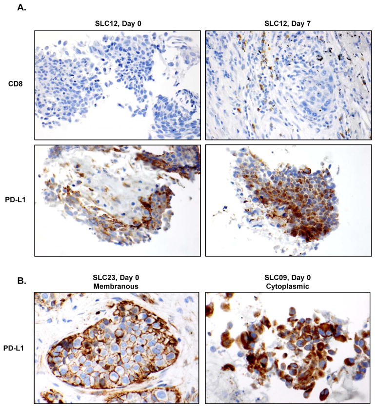
Figure 5. Tumor immunohistochemical staining with CD8 and PD-L1.
(A) Paraffin-embedded tumor tissues were stained with anti-CD8 and anti-PD-L1 on day 0 and day 7 and representative immunohistochemical staining images from SLC12 are shown. (B) Representative immunohistochemical staining images of membranous and cytoplasmic expression of PD-L1 on day 0 from patients SLC23 and SLC 09, respectively, are shown.

