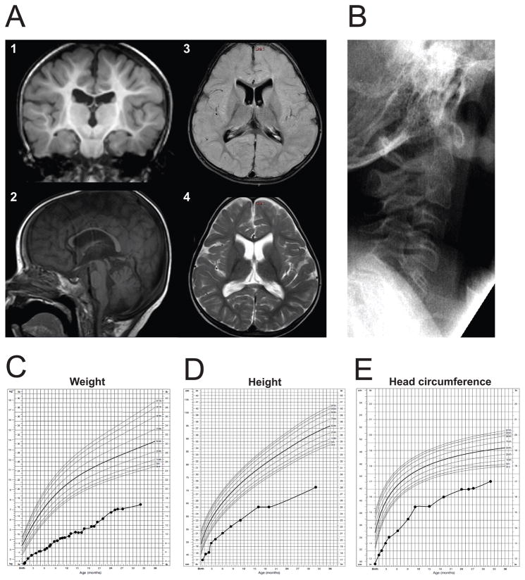
Figure 1.
Clinical features of patient UDP5316. (A) Representative MRI images for the proband at 29 months of age. Cerebral cortex appeared normal, but there was decreased cerebral white matter volume and delayed myelination (shown in 1, coronal image, T1-weighted), moderate-to-severe thinning of the corpus callosum, mild atrophy of the cerebellar vermis, small brain stem (shown in 2, sagittal image, T1-weighted), and mildly enlarged ventricles (axial images; 3, FLAIR; 4, T2-weighted). (B) Cervical spine X-ray shows fusion of C5–C7 vertebra. (C–E) Growth curves of the patient plotted in comparison with normal children 0–36 months of age; weight (C), height (D), and head circumference (E).

