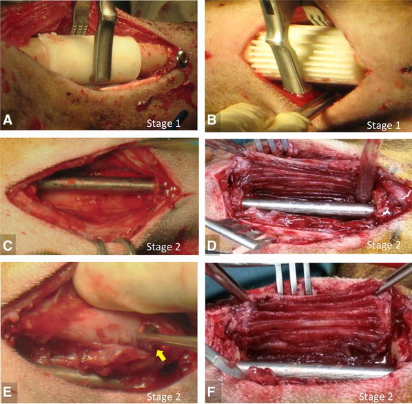
Fig. 1A–F.

The intraoperative photographs show placement of a (A) smooth spacer and (B) textured spacer in the defect site during the “spacer procedure” (Stage 1, Masquelet technique). Four weeks later during the “grafting procedure” (Stage 2, Masquelet technique), the induced membrane is appreciated after removal of the (C) smooth and (D) textured spacers. Scraping the inner layer of the induced membrane with a curette (yellow arrow) was done in half of the goats from the (E) smooth and (F) textured spacer groups.
