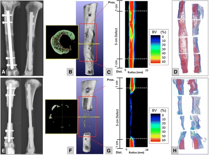
Fig. 5A–H.

Examples of two representative study goats are presented. One goat showed robust bone formation on (A) postmortem AP and lateral view, (B) micro-CT 3-D reconstruction and axial image (yellow box), (C) % bone volume (BV) plot versus summation of radial position, and on (D) Masson’s trichrome-stained histology slides (pink staining shows substantial bone formation). And another goat showed minimal new bone formation on (E) postmortem AP amd lateral radiographs, (F) micro-CT 3-D reconstruction and axial image (yellow box), (G), % bone volume (BV) plot versus summation of radial position, and on (H) Masson’s trichrome-stained histology slides (pink staining shows substantial bone formation).
