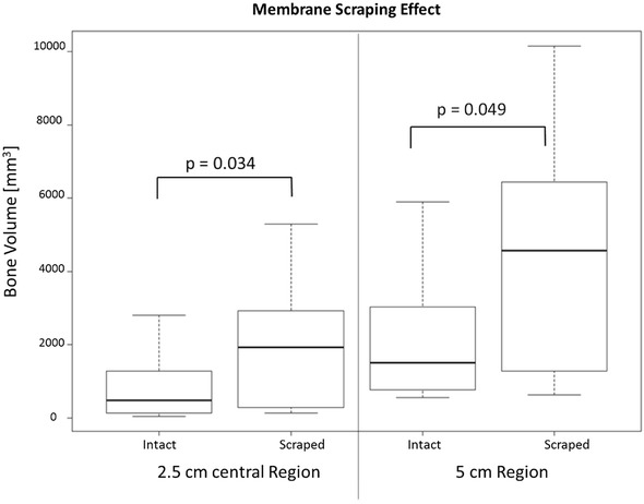
Fig. 6A–B.

The box plots show the distribution of total bone volume (based on micro-CT analyses) based on intact membrane or membrane scraped in the (A) central 2.5 cm of the defect (left) and the (B) entire 5-cm defect (right). The scraped induced-membrane group showed higher total bone volume compared with the intact membrane group in the 2.5 cm region (p = 0.034), confirming the hypothesis based on the primary outcome parameter. Post hoc (nonmodel based) comparison of tBV in the entire 5 cm defect using a one tailed t test found p = 0.049.
