Fig. 1.

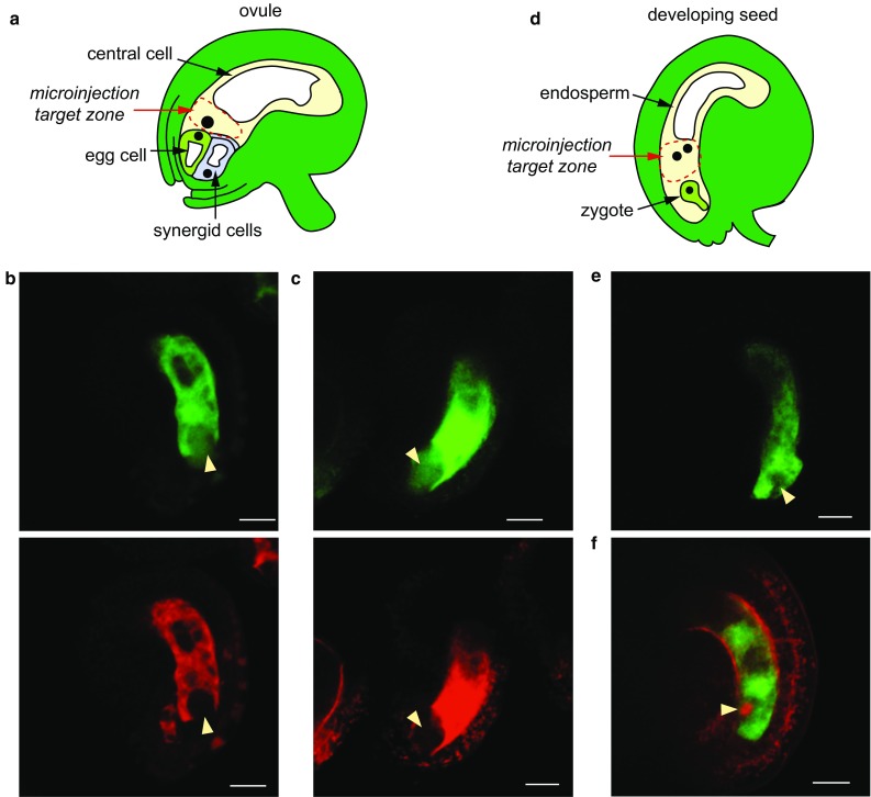
Assaying passive dye permeability in the female gametophyte before and after fertilization. a Schematic of Arabidopsis ovule. b Pre-fertilization co-injection of Ler with 10 kDa FITC (green) and 70 kDa rhodamine (red). Image captured 45 min following injection. c Pre-fertilization co-injection of Ler with 20 kDa FITC (green) and 70 kDa rhodamine. Image captured 80 min following injection. Yellow arrowheads indicate the position of the egg apparatus (the cluster of the egg cell and synergid cells). d Schematic of the developing seed, about 18 h after pollination. e Post-fertilization injection of Ler with 10 kDa FITC. Image captured 90 min after injection. f Post-fertilization injection of Ler FGR7.0 with 10 kDa FITC. RFP (red) is expressed within the zygote. Image captured 60 min after injection. Yellow arrowheads mark the position of the zygote. All scale bars, 20 μm
