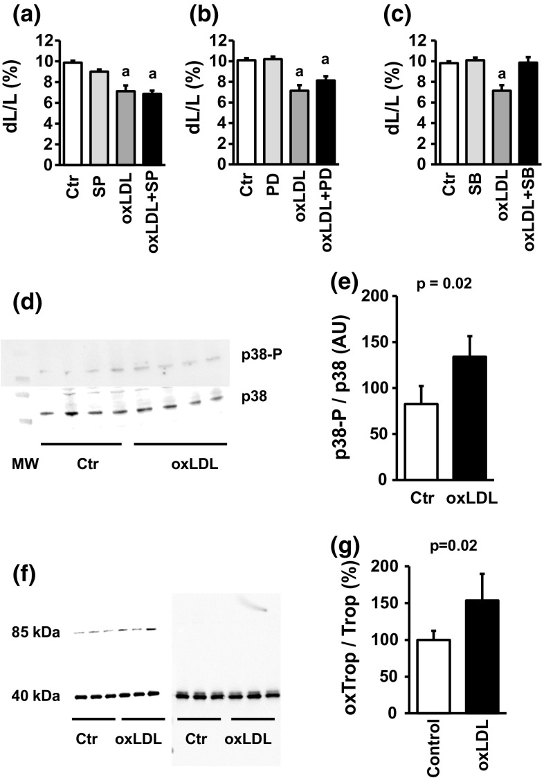
Fig. 4.
Participation of MAPK pathways in oxLDL-dependent effects: a cell shortening of cardiomyocytes exposed to oxLDL (20 µg/ml) and SP600125 (10 µM). b Cell shortening of cardiomyocytes exposed to oxLDL (20 µg/ml) and PD98059 (10 µM). c Cell shortening of cardiomyocytes exposed to oxLDL (20 µg/ml) and SB202190 (10 µM). In a–c a represents p < 0.095 vs. control. d Representative immunoblot of samples from cardiomyocytes exposed to oxLDL (20 µg/ml) and quantified for p38 MAPK expression and phosphorylation of p38 MAPK. e Quantification of the blot shown in d. f Original western blot showing the oxidative modification of tropomyosin by oxLDL (20 mg/ml) in the left and the control blot under reducing conditions (right). g Quantification of oxidative modification of tropomyosin by oxLDL (n = 3). Exact p values are given

