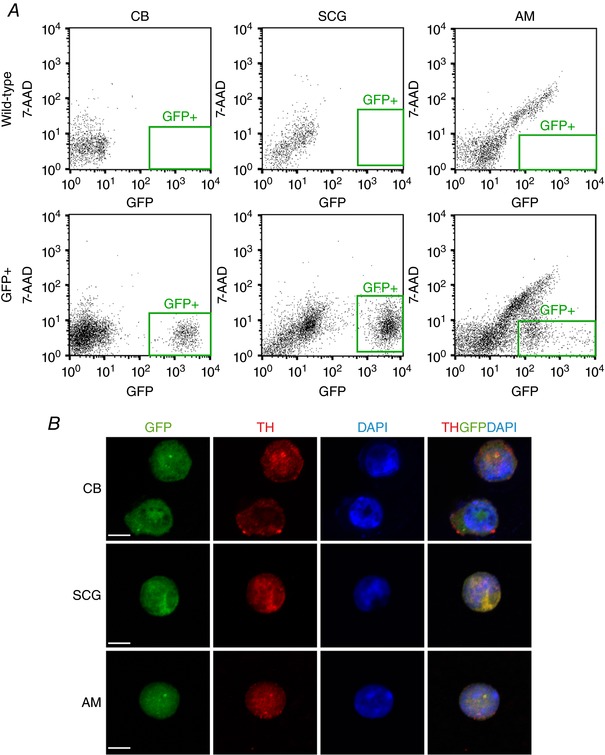
Figure 2. Sorting of tyrosine hydroxylase (TH) positive cells from adult wild‐type and TH‐GFP mice by flow cytometry.

A, isolation of green fluorescent protein positive (GFP+) cells from the carotid body (CB), superior cervical ganglion (SCG) and adrenal medulla (AM) of TH‐GFP mice by flow cytometry. 7‐AAD, 7‐aminoactinomycin D. Plots from wild‐type animals are shown as control. B, immunofluorescent staining demonstrating that the GFP+ cells express TH. Scale bar, 5 μm.
