Fig. 3.

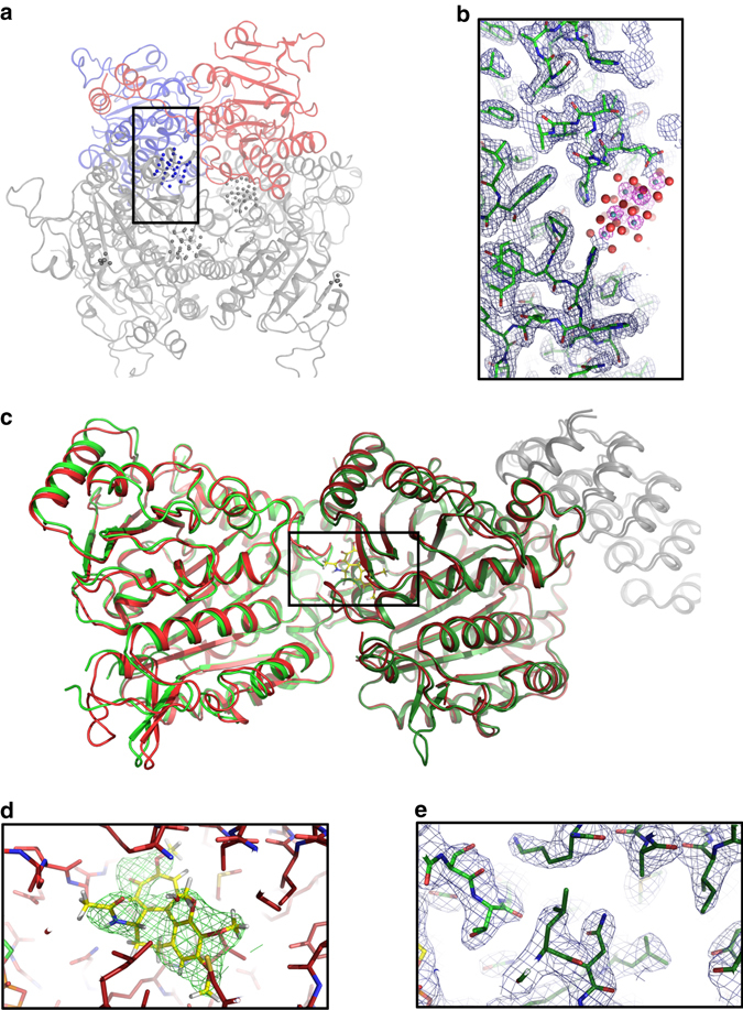
Structures of the molybdenum storage protein (MOSTO) and Tubulin (TD1). a The protein forms a heterohexameric (αβ)3 cage-like structure with polyoxomolybdate clusters bound inside the cage. The α- and β- subunits present in the asymmetric unit of the SMX structure are shown as red and blue cartoons and the remaining two αβ-units completing the heterohexamer are shown in gray. b The zoomed polyoxomolybdate cluster binding site was drawn for the polypeptide with an 2Fo–Fc electron density in blue at 1σ and for the molybdenum cluster (molybdenum and oxygen shown as gray and red spheres) in pink at 10σ, highlighting the positions of the polynuclear molybdenum-oxide aggregates. c Overlay of TD1apo and TD1col SMX structures shown as green and red cartoons, respectively. The DARPin is shown as gray cartoon and α-tubulin and β-tubulin subunits are shown as bright red or bright green and dark red or dark green, respectively. d Zoom in on TD1col. Surrounding residues are shown as red sticks and colchicine is shown as yellow sticks with the surrounding Fo–Fc density of the structure refined without the ligand at 3σ. e Zoom in on TD1apo. Surrounding residues are shown as green sticks with the surrounding 2Fo–Fc density of the structure at 1σ
