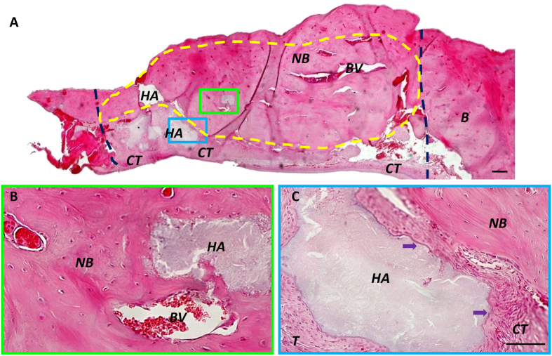
Figure 6.
HA/TCP mediated moderate new bone formation in mandibular bone defects. (A) Two months after surgery, the control group treated with HA/TCP alone showed moderate amounts of new bone in the defects. HA/TCP was also detected in the mineralized tissues (dark blue lines show the margins of the bone defect made in surgery). (B) Higher magnification of the area enclosed in a green rectangle in (A) shows newly formed bone was detected around HA/TCP particles. There were abundant blood vessels in the newly formed bone. (C) Control HA/TCP specimens show large volumes of HA/TCP particles remaining in the bone defect area, around connective tissues (purple arrows; the area enclosed with a blue rectangle in (A). HA = HA/TCP particles, NB = new bone, CT = connective tissue, BV = blood vessel. Scale bar = 200 µm in (A) and Scale bar = 50 µm in (B,C).

