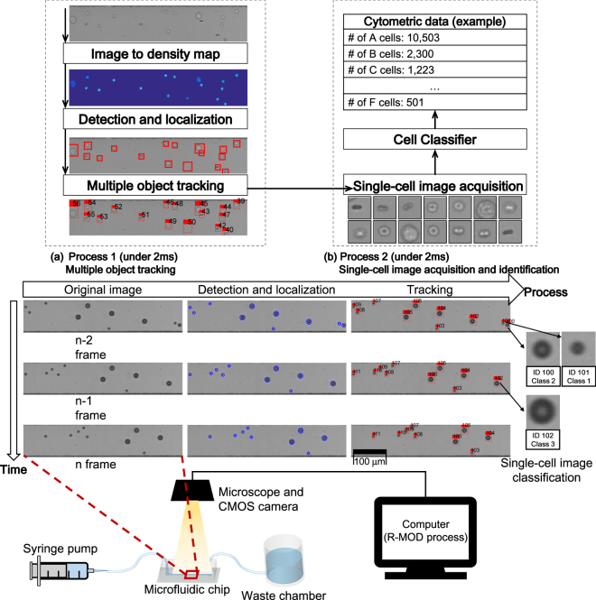
Figure 1.
Schematic of the designed system and R-MOD. CMOS camera on microscope observes the microfluidic channel through which cell suspension flows. Bright-field microscopic image taken by the CMOS camera is represented by a grayscale image. The resolution of the image is 100 by 500 pixels and the CMOS camera collects image sequences at 500 frame/s (i.e. every 2 ms). Thus, R-MOD must perform the whole process in <2 ms. Two processes are executed in parallel (multi-threads) and each process should perform its task <2 ms. (a) Process 1 performs detection/localization and multiple object tracking. For the detection and localization tasks, FCRN converts the original grayscale images to probability density maps (<1.5 ms), then the Flattening algorithm extracts centre positions and sizes from the maps (<0.1 ms). After obtaining location of each cell, the multiple-object tracking algorithm finds correspondences between consecutive frames (<0.2 ms); this step eliminates repeated counting of cells in flow. (b) Process 2 performs single-cell image acquisition and identification. Using the tracking result, single-cell images can be obtained by cropping them from the original ROI image frame without duplications. The cropped single-cell images are evaluated by an image classifier based on a supervised learning to identify cell type.

