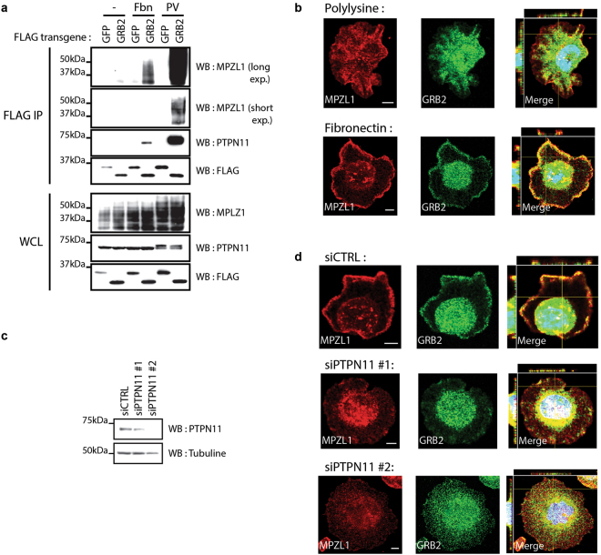
Figure 4.
Cell adhesion on fibronectin triggers GRB2-MPZL1 complex formation at the plasma membrane. (a) HEK293T cells expressing FLAG-GFP or GRB2 were seeded on plastic or fibronectin for ten minutes before lysis. PV treated cells were used as a positive control. FLAG APs were performed for each condition and analyzed by Western blot as indicated. (b) HCC1954 cells were seeded on polylysine or fibronectin for 15 minutes before fixation. Immunofluorescence against GRB2 and MPZL1 was performed to assess the cellular localization. Confocal images of the z-axes (indicated by yellow lines) are shown on the left side (y-z axis) or on top (x-z axis) of the panels showing merged images, for which nuclei were stained with DAPI. Representative images are presented (scale bar = 5 μm). (c) HCC1954 cells were depleted of endogenous PTPN11 using DsiRNAs. (d) PTPN11-depleted cells were seeded on fibronectin for 15 minutes prior to fixation and immunofluorescence. Confocal images of the z-axes (indicated by yellow lines) are shown on the left side (y-z axis) or on top (x-z axis) of the panels showing merged images, for which nuclei were stained with DAPI. Representative images are shown (scale bar = 5 μm).

