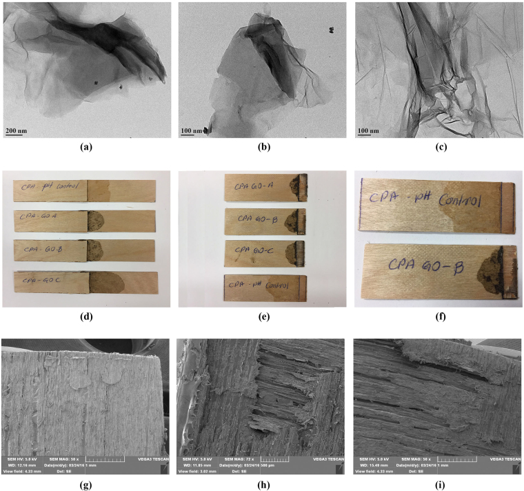
Figure 7.
Image (a–c) represent the transmission electron microscopic (TEM) images of exfoliated graphite oxide samples prepared under different oxidation time in canola protein matrix. (a) CPA GO-A, (b) CPA GO-B and (c) CPA GO-C are prepared by exfoliating 1% w/w (GO/protein) GO-A, GO-B and GO-C respectively in 10% w/v canola protein dispersion. (d–f) represent the images of wood veneer samples bonded with CPA GO adhesives (d), and their fracture surface after strength testing (e,f). Images (g–i) represent the scanning electron microscopic (SEM) images of wood fracture surface after bond testing for (g) CPA GO-A adhesive, (h) CPA GO-B adhesive and (i) CPA GO-C adhesive

