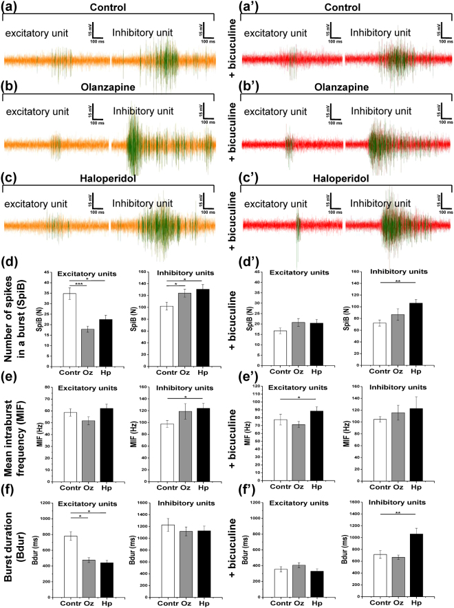
Figure 5.
Olanzapine (Oz) and haloperidol (Hp) differentially modify burst structure in excitatory and inhibitory units. The activity line plots and quantifications are shown for 21 days in in vitro cultures. (a–c) The representative bursts are shown for putative excitatory and inhibitory units in control, olanzapine, and haloperidol conditions, respectively. (a’–c’) The bursts in the same units as in (a–c) are shown after 30 minutes of 6 µM bicuculline methiodide application. Vertical bar, 15 mV; horizontal bar, 100 ms. (d–f) The quantification of key burst structure parameters is shown for putative excitatory units (minimum 115 electrodes, N = 4) and inhibitory units (minimum 32 electrodes, N = 4): number of spikes in a burst (SpiB) (d) mean intraburst spiking frequency (MIF) (e) and burst duration (Bdur) (f). (d’–f’) Burst structure parameters are quantified after 30 minutes of 6 µM bicuculline methiodide application, same units as in (d–f). The asterisks indicate significant differences with the control, based on the Kruskal-Wallis ANOVA test (*p < 0.05; **p < 0.01; ***p < 0.001).

