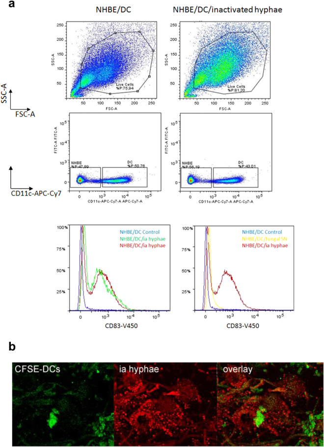
Figure 5.
DC maturation and activation upon fungal exposure of the epithelia. (a) DC maturation is induced by inactivated (ia) hyphae. Gating of non-infected (upper, left) and A. fumigatus-infected (upper, right) NHBE/DC co-cultures revealed that under both conditions about 80% were alive of which ~50% showed a high expression of the characteristic DC marker CD11c, while the other half did not express this marker, thereby characterizing these CD11c-negative cells as NHBE cells (middle panel). The CD11chigh population of non-infected and infected co-cultures was further characterized for the DC maturation marker CD83. Only upon apical application of inactivated A. fumigatus hyphae a significant up-regulation of CD83 was detectable as shown on cells harvested from two independent Transwells (lower panel, left, red and green), while control cells did not mature (lower panel, left, blue). Also the application of fungal SNs to the apical surface of the epithelium (lower panel, right, yellow) did not cause DC maturation as high as inactivated A. fumigatus hyphae (lower panel, right, red). Compared to uninfected control cells (lower panel, right, blue), only a slight shift to the right was observable respecting CD83 upon treatment of the epithelium with fungal SNs (lower panel, right, yellow). FACS analyses were repeated thrice. (b) CLSM analyses revealed apical DC attachment to ia hyphae. To see if PAMPs are sufficient for DC attraction, apical epithelial layers were exposed to ia hyphae (red) for 48 h. CFSE-DCs (green) showed a co-localization with hyphae (overlay). CLSM analyses using ia hyphae and counting at least 25 cells were performed twice.

