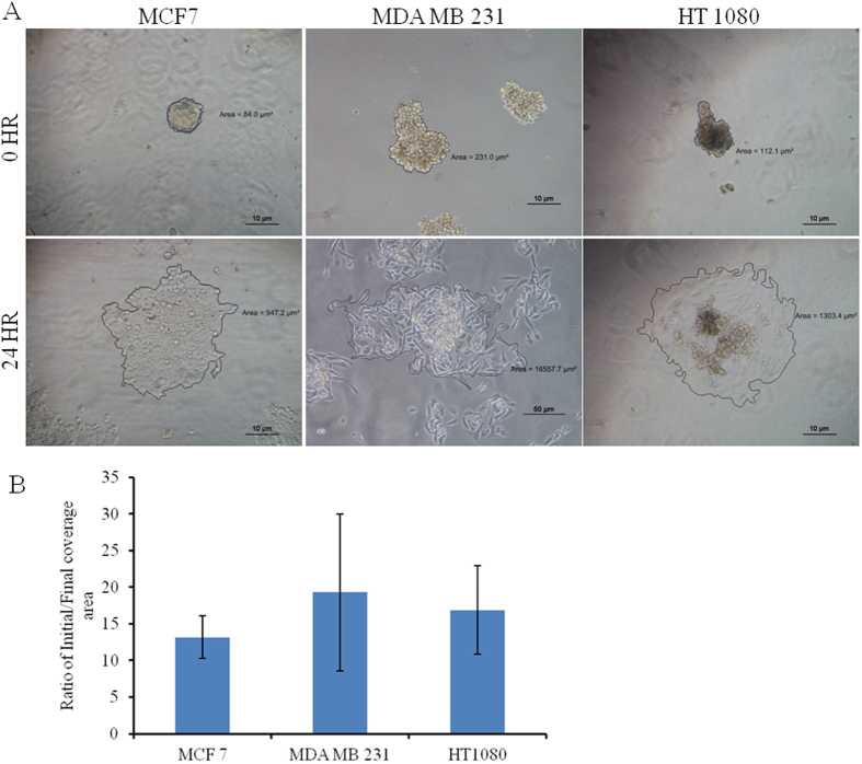
Figure 3.
Pseudo 3D migration of MCF7, MDA MB 231 and HT 1080 spheroid on the 2D surface. (A) A single spheroid is incubated on the glass coverslip and incubated for 24 hr. Reversal /melting of spheroids over time is analyzed by imaging with phase contrast mode of Nikon Eclipse Ti-U. The scale bar is 10 µm. (B) The covered area is calculated (NIS Elements software) and normalized with initial spheroid size for plotting (mean ± SEM).

