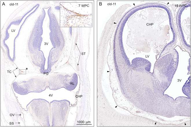
Figure 5.
Claudin-11 immunoreactivity in a 7 wpc embryo and a 10 wpc fetus. (A) 7 wpc embryo: the arachnoid barrier cell layer appears initially covering the inner surface of the tentorium cerebelli (TC); it is most prominent in the medial part where the ‘blades’ of the tentorium meet (insert). Note that there is no indication of a claudin-11 positive arachnoid layer covering the forebrain (A). The choroid plexus (CHP) of the 4th ventricle (4 V), positively immunostained for claudin-11, is starting to differentiate, and the sinuses are already present. (B) 10 wpc fetus: the basal forebrain shows a strongly claudin-11 immunopositive arachnoid barrier cell layer (four basolateral arrowheads) creating a well-defined subarachnoidal space in contrast to the dorso-medial forebrain where the claudin-11 immunopositive arachnoid membrane fades out along the surface (three upper lateral arrowheads). Abbreviations: CHP: choroid plexus; 4 V: fourth ventricle; LV: lateral ventricle; OV: otic vesicle; PG: pineal gland; RL: rhombic lip; 3 V: third ventricle; SS: sigmoid sinus; ST: transverse sinus; TC: tentorium cerebelli; WPC: weeks post conception; cld: claudin. (A,B) Same magnification. Scale bar: 1000 μm.

