FIG 2.
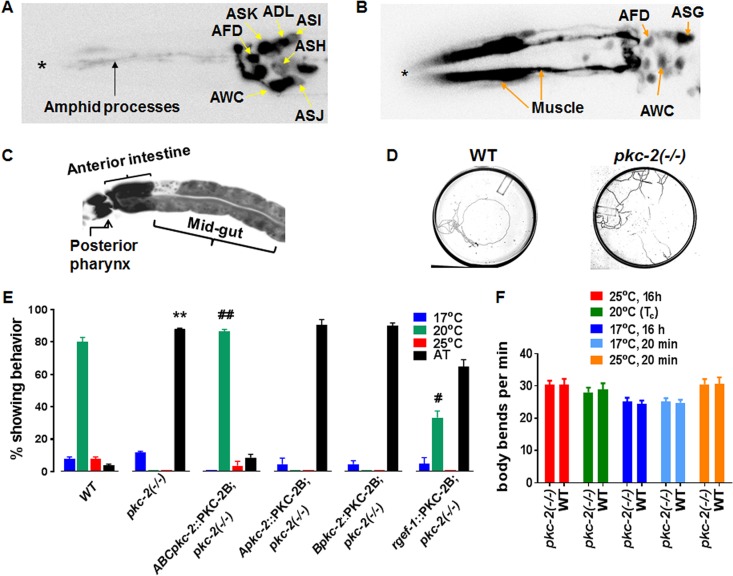
pkc-2 promoters are active in sensory neurons, muscle, and intestine; PKC-2 depletion disrupts thermotaxis. (A) Neurons engaged in pkc-2 gene transcription were identified by fluorescence microscopy of animals expressing GFP under the control of the pkc-2 A promoter (Apkc-2::GFP). The micrograph extends from the head (left, asterisk) to the top of the gut (right). (B) Animals expressing a Bpkc-2::GFP transgene were analyzed as described for panel A. (C) Fluorescence microscopy also revealed pkc-2 B promoter activity in pharyngeal neurons and intestine. (D) Tracks produced when WT and pkc-2−/− animals experienced a radial temperature gradient (17°C [center] to 25°C [periphery]) on a 10-cm agar plate. The Tc was 20°C. (E) WT, PKC-2-deficient, and the indicated transgenic animals were assayed for thermotaxis behavior (see Materials and Methods). Percentages of animals that migrated to the center (17°C), the Tc (20°C), or the periphery (25°C) or exhibited AT behavior were determined. Thermotaxis assays were performed with WT C. elegans, pkc-2−/− animals, and pkc-2−/− animals expressing PKC-2B transgenes controlled by all three pkc-2 promoters (ABCpkc-2::PKC-2), the pkc-2 A promoter, pkc-2 B promoter, or a pan-neuronal rgef-1 promoter (32). Results are averages obtained from three assays. Error bars are SEM. **, P < 0.01 compared to WT animals; ##, P < 0.01 compared to pkc-2−/− animals; #, P < 0.05 compared to pkc-2−/− animals. (F) WT and pkc-2−/− animals were assayed for locomotion on a bacterial lawn (see Materials and Methods) after brief (20 min) or prolonged (16 h) incubation at 17°C, 20°C, or 25°C. The number of body bends in a 1-min interval was determined for 10 WT or pkc-2−/− animals. Mean values and SEM are shown.

