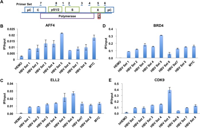
FIG 1.
Different P-TEFb-containing complexes are recruited to the HBV genome. (A) Cartoon model illustrating the position of the primer pairs used for qPCR after ChIP along the HBV genome. (B to E) The SEC subunits AFF4 (B), ELL2 (C), and BRD4 (D) and the P-TEFb kinase module CDK9 (E) are all recruited to the HBV genome, as detected in HepG2.2.15 cells by ChIP-qPCR assays with specific antibodies. The HEMO gene serves as a negative control for ChIP-qPCR. Error bars represent the standard deviations from three independent measurements.

