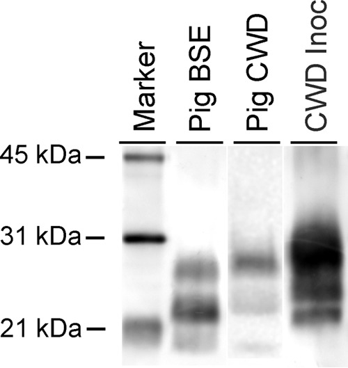
FIG 1.

WB analysis demonstrating a unique PrPSc profile in brain samples from pigs with CWD. The positive brain sample from a pig inoculated with the CWD agent (pig CWD) has a slightly higher migration than the brain sample from a pig inoculated with the agent of classical BSE (pig BSE) and a much lower migration than the CWD inoculum (CWD Inoc). The diglycosylated band (topmost band in each lane) is more prominent in the pig CWD and CWD Inoc samples, while the monoglycosylated (middle) band is most prominent in the pig BSE sample. The blot was developed with monoclonal antibody L42. Note that because of the sparse PrPSc accumulation in the brains of inoculated pigs, the blot shown is a composite; see Materials and methods for details.
