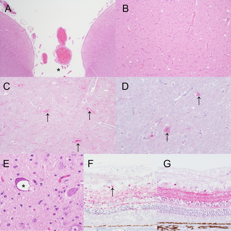
FIG 4.
Vacuolar change and PrPSc in the brain and eye. (A) Brain stem of pig 7 showing incidental, i.e., unrelated to prion disease, neuropil vacuolation in the colliculus. *, midline (hematoxylin and eosin staining; original magnification, ×4). (B) Higher-magnification view of panel A (original magnification, ×10). (C) Brain stem of pig 25 showing intraneuronal PrPSc immunoreactivity (arrows) in neurons in the colliculus (anti-PrP monoclonal antibody L42; original magnification, ×20). (D) Brain stem of noninoculated control pig 8 showing non-disease-specific intraneuronal immunolabeling (arrows) in neurons in the colliculus (anti-PrP monoclonal antibody L42; original magnification, ×40). (E) Brain stem of pig 38 showing incidental intraneuronal vacuolation (*) in the dorsal motor nucleus of the vagus nerve (hematoxylin and eosin staining; original magnification, ×40). (F) Retina of pig 26 showing granular to punctate PrPSc immunoreactivity in the inner and outer plexiform layers with occasional intraglial deposits (arrow) (anti-PrP monoclonal antibody L42; original magnification, ×40). (G) Retina of noninoculated control pig 4 showing non-disease-specific immunolabeling (anti-PrP monoclonal antibody L42; original magnification, ×40).

