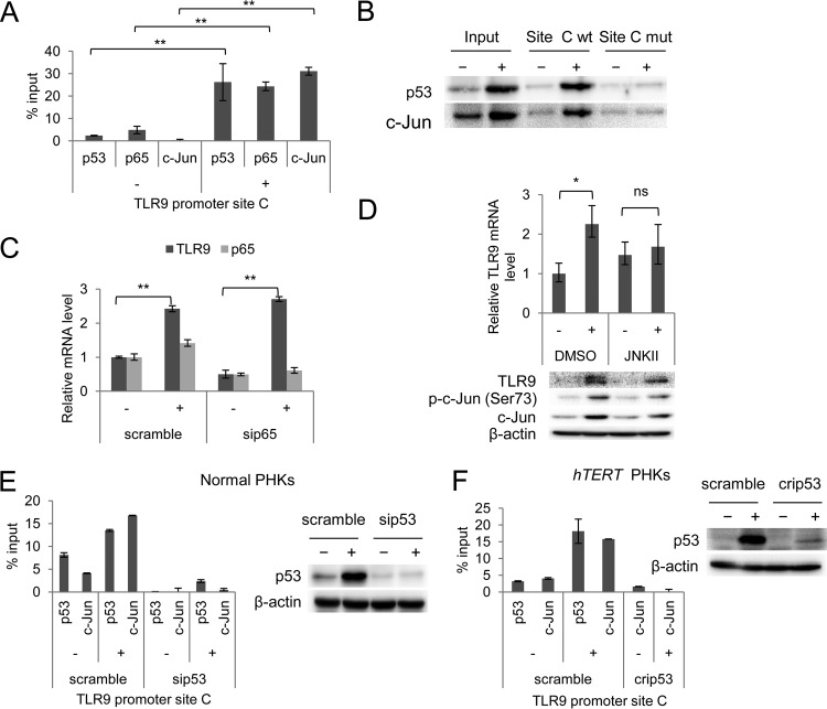
FIG 5.
UV-induced TLR9 transcriptional activation is mediated by p53 and c-Jun recruitment at a specific region of the TLR9 promoter. (A) ChIP was performed in nonirradiated (−) and UVB-irradiated (+) normal PHKs using p53, p65, and c-Jun antibodies. Simultaneously, part of the total chromatin fraction (1/10) was used as the input. qPCR was performed using specific primers flanking NF-κB RE C within the TLR9 promoter. The histogram shows the relative amount of the promoter bound by the antibodies after subtracting the background of nonspecific IgG control, expressed as a percentage of the input. The data shown are representative of three independent experiments. (B) The UVB-irradiated (+) and nonirradiated (−) hTERT PHKs were processed for the oligonucleotide pulldown assay. Cell lysate was incubated with biotinylated probes containing NF-κB RE C of the TLR9 promoter, either wild-type (site C wt) or mutated (site C mut). DNA-associated proteins were recovered by precipitation with streptavidin beads and analyzed by immunoblotting. (C) Normal PHKs were transfected with a control siRNA (scramble) or an siRNA specific for p65 (sip65). After 40 h, the cells were UVB irradiated (+) or mock irradiated (−) as described in Materials and Methods. The total RNA was then extracted, and p65 and TLR9 mRNA levels were measured by RT-qPCR. The error bars represent the standard deviations of two biological replicates. (D) Normal PHKs were treated with either JNK inhibitor II (JNKII) or dimethyl sulfoxide (DMSO) and then UVB irradiated (+) or mock irradiated (−) as described in Materials and Methods. After 8 h, the total RNA was extracted, and the TLR9 mRNA levels were measured by RT-qPCR (upper panel). Simultaneously, total proteins were extracted and analyzed by immunoblotting for the indicated antibodies (lower panel). The error bars represent the standard deviations of two biological replicates. (E) Normal PHKs were transfected with a control siRNA (scramble) or an siRNA specific for p53 (sip53). After 40 h, the cells were UVB irradiated (+) or mock irradiated (−) as described in Materials and Methods; total cellular extracts were processed for ChIP with the indicated antibodies. (F) hTERT PHKs either expressing wild-type p53 (scramble) or with CRISPR/Cas9-mediated p53 deletion (crip53) were UVB irradiated (+) or mock irradiated (−) and then processed for ChIP with the indicated antibodies. *, P < 0.05; **, P < 0.01; ns, not significant.

