FIG 1.
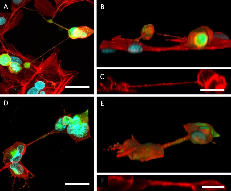
US3-induced cell projections are tunneling nanotubes (TNTs). (A to C) Confocal image of cell projections in US3-transfected ST cells showing GFP cotransfected with US3 (green), filamentous F-actin (red), and nuclei (cyan). (A) Maximum projection image of different xy optical sections through the sample, showing the presence of F-actin in US3-induced cell projections. Bar, 30 μm. (B and C) Three-dimensional (3D) reconstruction (B) and xy section (C) of the same confocal image, showing that the US3-induced cell projections do not make contact with the underlying substrate. Bar, 20 μm. (D to F) Confocal image of cell projections in PRV-infected ST cells showing viral gD protein (green), filamentous F-actin (red), and nuclei (cyan). (D) Maximum projection image of different xy optical sections through the sample, showing the presence of F-actin in PRV-induced cell projections. Bar, 30 μm. (E and F) 3D reconstruction (E) and xz section (F) of the same confocal image, showing that the PRV-induced cell projections do not make contact with the underlying substrate. Bar, 20 μm.

