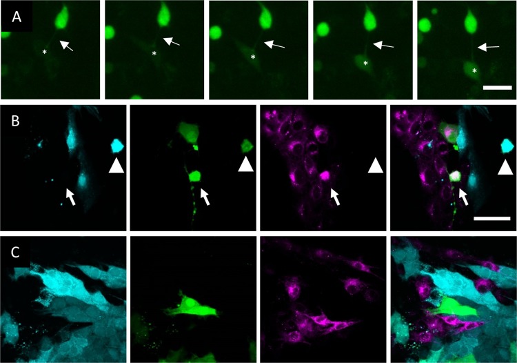
FIG 2.
US3-induced cell projections allow transport of GFP signal from transfected to untransfected cells. (A) GFP signal is transferred from US3/GFP-cotransfected ST cells to nontransfected ST cells. Arrows show a TNT connecting a US3/GFP-transfected cell to a nontransfected cell. Asterisks show the location of an acceptor cell that becomes GFP positive over time. The interval between each frame and the next is 30 min. Bar, 30 μm. Images represent stills from a time lapse video (Movie S2). (B) Coculture experiment results showing transfer of GFP (green) from a wild-type US3/GFP-transfected CellTrace violet-stained (cyan) donor population (arrowhead) to a nontransfected Did-stained (magenta) accepter population (arrow). (C) Use of the same experimental setup as that described for panel B but using kinase-inactive US3 instead of wild-type US3 did not lead to GFP transfer to nontransfected cells. Bar (for panels B and C), 50 μm.

