FIG 4.
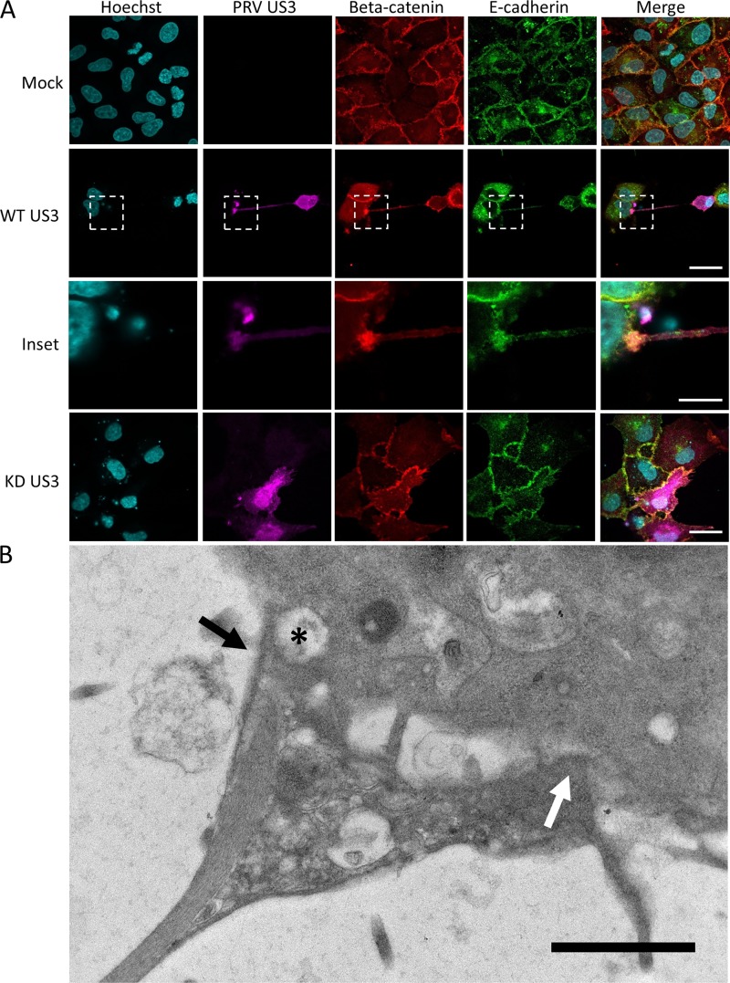
The contact area between US3-induced TNTs and acceptor cells is enriched in E-cadherin and beta-catenin in transfected cells and has a heterogeneous structure. (A) E-cadherin (green) and beta-catenin (red) staining in US3-transfected (magenta) RK13 cells. Bars, 30 μm; inset bar, 10 μm. (B) Transmission electron microscopy (TEM) image of an area of contact between a TNT and acceptor cell in US3-transfected ST cells. An area with loose contact is indicated with a white arrow, while an area with apparent cytoplasmic connectivity between the two cells is indicated with a black arrow. A vesicle appears to be transported through this region with cytoplasmic connectivity (black asterisk). Bar, 1 μm. KD, kinase dead; WT, wild type.

