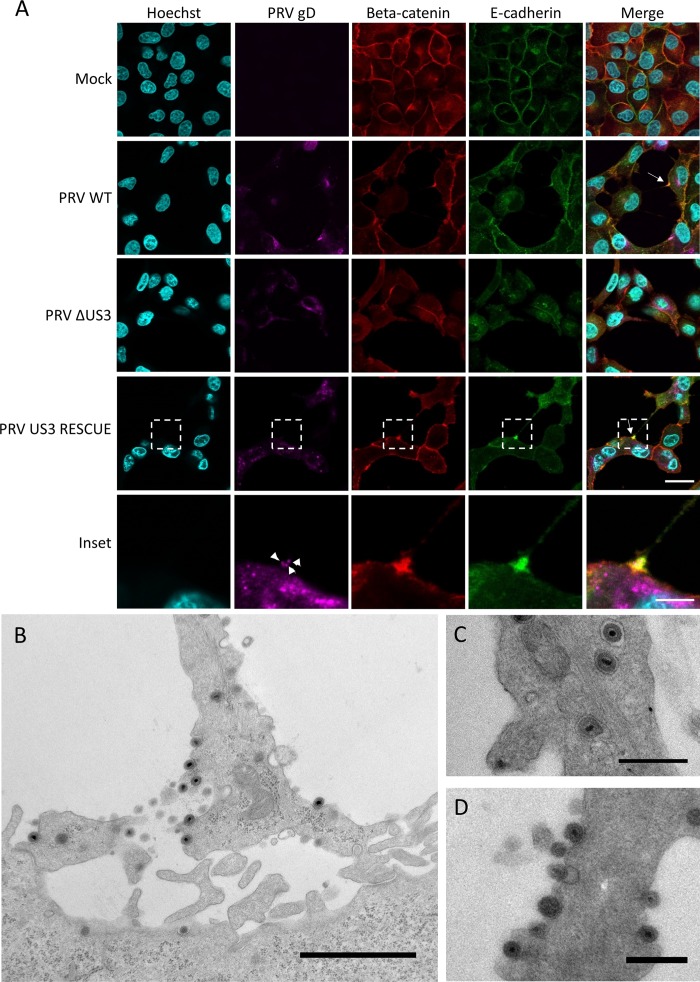
FIG 5.
The contact area between US3-induced TNTs and acceptor cells in PRV-infected samples is enriched in E-cadherin and beta-catenin, and enveloped virus is present in vesicles in the TNTs and is released along the length of the TNT and at the contact zone. (A) E-cadherin (green) and beta-catenin (red) staining in PRV-infected (magenta) RK13 cells. White arrows show areas enriched in beta-catenin and E-cadherin. Arrowheads indicate punctate gD signal in the contact zone, indicating the presence of virus particles. Bars, 30 μm; inset bar, 10 μm. (B to D) Transmission electron microscopy (TEM) images of ST cells infected with wild-type PRV. (B) Contact area of a US3-induced TNT with a nearby cell in ST cells infected with PRV. Bar, 1,500 nm. (C to D) Virus particles are transported through TNTs as individually membrane-wrapped enveloped virions and are released over the entire length of the conduit. Bars, 500 nm. WT, wild type; PRV, pseudorabies virus; ΔUS3, US3null mutant.

