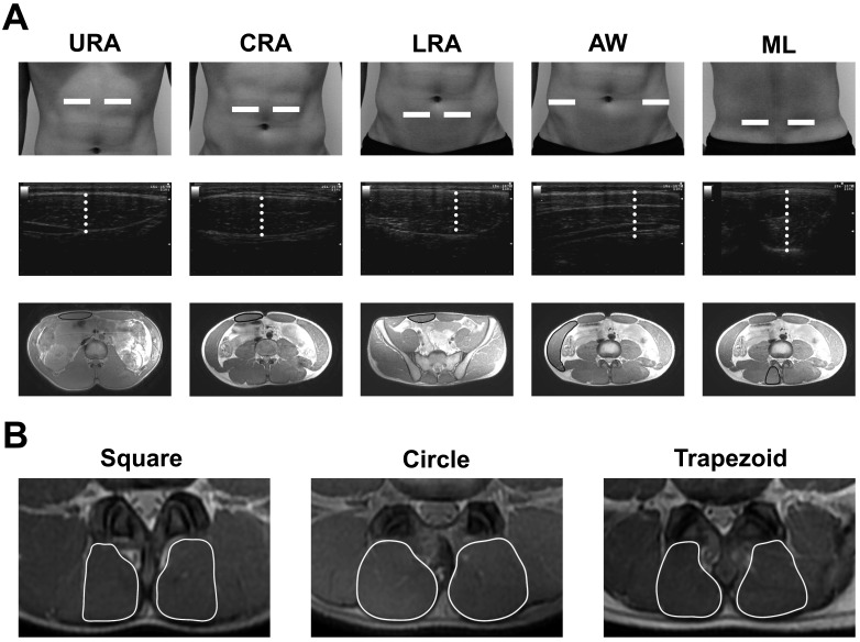
Fig. 1.
Representative images of ultrasonography (US)-measured muscle thickness (MT) and magnetic resonance imaging (MRI)-measured muscle cross-sectional area (MCSA) of the trunk muscles. Panel A shows the probe positions for the US measurements (upper panel) and representative images of US-measured MT (center panel) and MRI-measured MCSA (lower panel) of the trunk muscles in a subject. The white dotted line in the US-measured MT image (center panel) denotes the MT. The black line in the MRI-measured MCSA image represents the MCSA. Panel B shows representative images of different forms of MCSA of the multifidus lumborum (ML) obtained from MRI measurements. URA: upper rectus abdominis; CRA: central rectus abdominis; LRA: lower rectus abdominis; AW: abdominal wall

