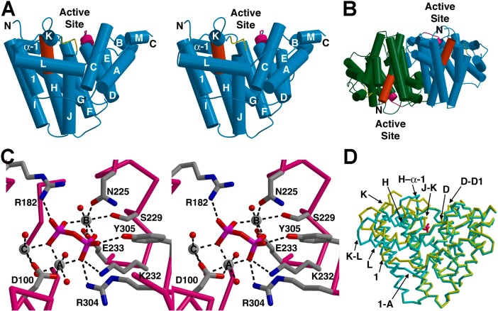
Figure 56.
(A) Stereoview of trichodiene synthase. The aspartate-rich motif on helix D (magenta) and the NSE motif on helix H (red) are indicated. (B) Two monomers assemble in antiparallel fashion to form the trichodiene synthase dimer. (C) Stereoview of the trichodiene synthase complex with inorganic pyrophosphate and 3 Mg2+ ions, showing metal coordination and hydrogen bond interactions (dashed lines). (D) Structure of unliganded trichodiene synthase (cyan) superimposed on that of the pyrophosphate complex (yellow, with magenta pyrophosphate) reveals structural changes in the indicated helices and loops that accompany active site closure. Reprinted from ref (170). Copyright 2001 National Academy of Sciences.

