
Figure 58.
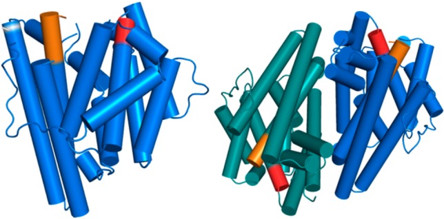
Structure of the monomer (left) and dimer (right) of germacradien-4-ol synthase; aspartate-rich and NSE metal-binding motifs are red and orange, respectively. The location of the E248A mutation required to facilitate crystallization is indicated by a white band. Reproduced from ref (279). Copyright 2016 American Chemical Society.