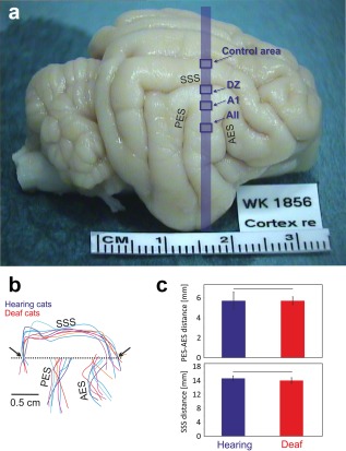
Figure 3.

Gross anatomy of the auditory brain is not changed in congenital deafness. (a) Brain of a congenitally deaf cat in toto. The blue stripe indicates the position of the evaluated sections: the blue rectangles indicate the position of the investigated cortical areas. (b) Schematic illustration of the sulcal patterns in the auditory cortex normalized in the dorsoventral direction with reference to the dorsal end of the PES (reference point), and in the rostrocaudal direction by the point where the rostrocaudal line crossing the reference point (dashed line) intersects with the caudal portion of the superior sylvian sulcus (arrow). (c) Analysis of the distance between AES and PES (top) and the distance between the caudal and rostral intersections of SSS with the dashed line marked by the arrows (b). All measurements were taken at the level indicated by the dashed line. None of the measures showed significant difference between hearing and congenitally deaf cats (α = 5%, two‐tailed Wilcoxon–Mann–Whitney test). AES: anterior ectosylvian sulcus; DZ: dorsal zone; A1: pimary auditory field; AII: secondary auditory field; PES: posterior ectosylvian sulcus; SSS: superior sylvian sulcus
