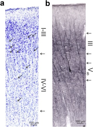
Figure 4.

Detail of the staining in a hearing control. (a) Nissl staining marks cells throughout the whole auditory cortex and reveals some aspects of cortical cytoarchitecture. Arrows in the image point to examples of pyramidal cells that are present in supragranular layers but not in layer IV; the arrows on the side point to the border of layer III and IV and the boundary between layer VI and white matter. (b) SMI‐32 stains somata and dendritic trees and reveals the dendritic anatomy in detail. The method stains cells in layer III and V. The cells are well differentiated from the background and indicate the cortical layer they are located in. The arrows on the side mark the borders of layer III and Vab as used in the present study to quantify layer thickness
