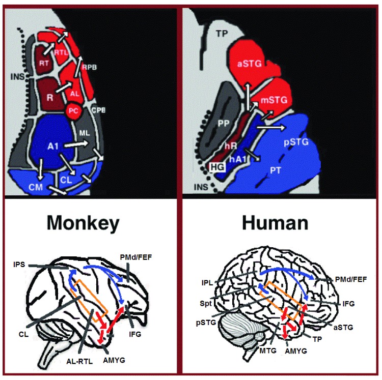
Figure 1. Dual stream connectivity between the auditory cortex and frontal lobe of monkeys and humans.
Top: The auditory cortex of the monkey (left) and human (right) is schematically depicted on the supratemporal plane and observed from above (with the parieto-frontal operculi removed). Bottom: The brain of the monkey (left) and human (right) is schematically depicted and displayed from the side. Orange frames mark the region of the auditory cortex, which is displayed in the top sub-figures. Top and Bottom: Blue colors mark regions affiliated with the ADS, and red colors mark regions affiliated with the AVS (dark red and blue regions mark the primary auditory fields). Abbreviations: AMYG-amygdala, HG-Heschl’s gyrus, FEF-frontal eye field, IFG-inferior frontal gyrus, INS-insula, IPS-intra parietal sulcus, MTG-middle temporal gyrus, PC-pitch center, PMd-dorsal premotor cortex, PP-planum polare, PT-planum temporale, TP-temporal pole, Spt-sylvian parietal-temporal, pSTG/mSTG/aSTG-posterior/middle/anterior superior temporal gyrus, CL/ML/AL/RTL-caudo-/middle-/antero-/rostrotemporal-lateral belt area, CPB/RPB-caudal/rostral parabelt fields.

