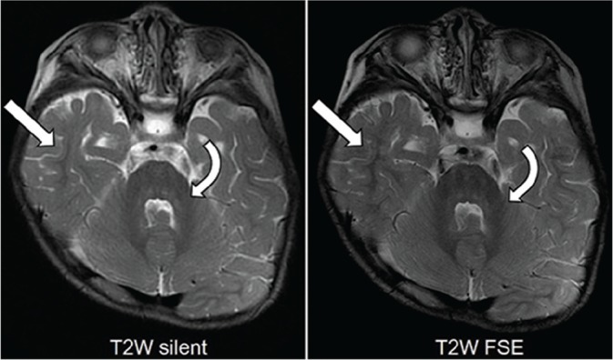
Fig 3.

T2-weighted (T2W) images from an 18-month-old boy with achondroplasia demonstrating low-signal myelin in the middle cerebellar peduncles (curved arrows), deep cerebellar white matter, and extending into the temporal subcortical white matter (arrows).
