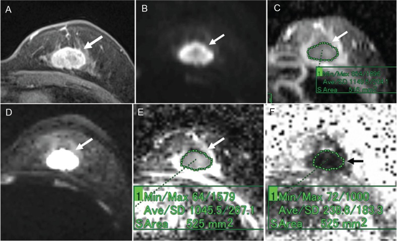
Fig 6.
A 59-year-old woman with invasive ductal carcinoma (triple negative cancer). (A) Post-contrast, fat-suppressed, axial T1-weighted image. (B) DWI based on ss-EPI. (C) ADC map of panel B. (D) DTI based on rs-EPI. (E) ADC map of panel D. (F) FA map of panel D. Post-contrast, fat-suppressed, axial T1-weighted image shows an oval-shaped mass with a slightly irregular margin and rim enhancement (A). The mass shows high signal intensity on both the DWI based on ss-EPI (B) and the DTI based on rs-EPI (D). On the ADC map of the DWI image, the ADC of the mass was 1.15 (C). On the ADC map of the DTI image, the ADC of the mass was 1.05 (E). On the FA map, the FA value of the mass was 0.24 (F).

