
Fig 4.
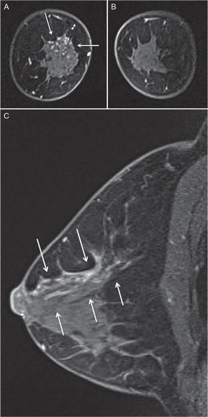
(A, B) Magnetic Resonance (MR) image of both breasts. Background parenchymal enhancement (BPE) is minimal. Coronal first contrast-enhanced fat-suppressed T1-weighted MR image showed segmental non-mass lesions (arrows) located at the upper portion of the right breast. (C) MR image of the right breast. Sagittal contrast-enhanced fat-suppressed T1-weighted MR image showed branching ductal patterns (arrows) located at the upper portion of the right breast.