
Fig 1.
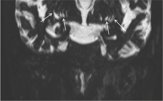
Coronal reformatting is performed to differentiate a large perivascular space (PVS) from the cerebrospinal fluid (CSF)-containing lacunes. On a coronal reformatted image, the PVS (arrows) shows a typical shape following perforating vessels without surrounding gliosis.