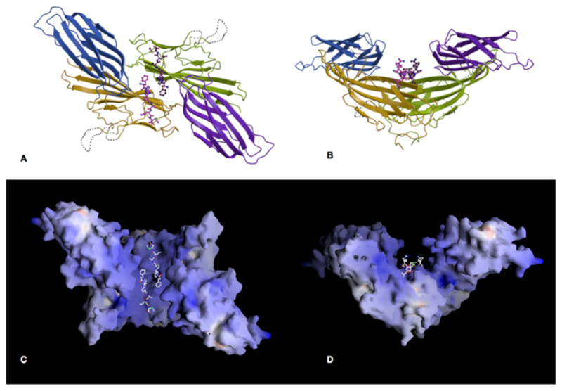
Figure 4. The crystallographic dimer.
A, B Orthogonal views of the dimer formed in the crystal, along and perpendicular to the crystallographic twofold axis. The A subdomains are coloured gold and green and the B domains blue and purple.
C and D. The surface of the μ2 dimer coloured according to electrostatic surface potential (blue positive, red negative, scale from -30 to +30 kT e-1)), in the same view as A and B. The planar face at the top of D may interact with the membrane. (Drawn with GRASP(33))

