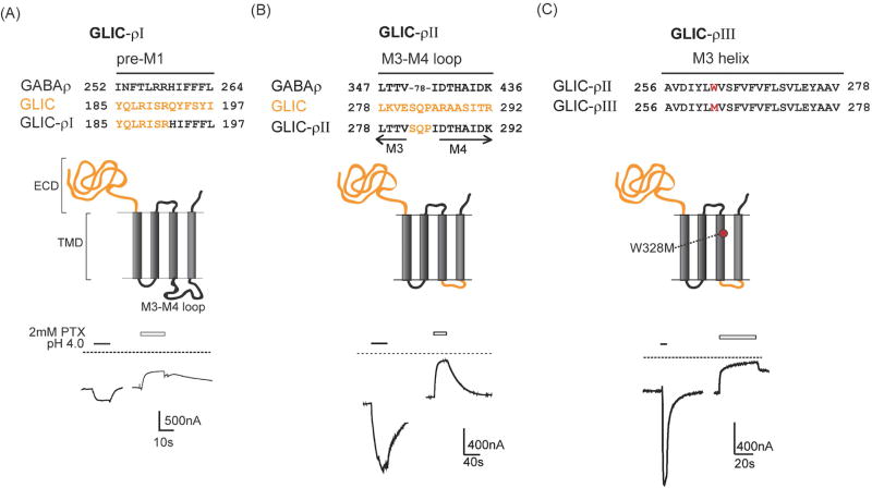
Figure 1. GLIC-GABAρ chimeric subunits form functional proton-gated channels.
(A) Top: Sequence alignments of pre-M1 regions of GABAρ, GLIC and GLIC-ρI chimera illustrating where ECD of GLIC was attached to TMD of GABAρ subunit; Middle: Schematic of GLIC–ρI chimeric subunit with GLIC protein in orange and GABAρ in black; Bottom: Representative pH 4.0 induced currents from an oocyte expressing GLIC–ρI chimeric channels. Dotted line represents zero current level highlighting resting leak current that is blocked by 2mM picrotoxin (PTX). (B) Top: Partial sequence alignments of M3, cytoplasmic M3–M4 loop and M4 of GABAρ, GLIC and GLIC-GABAρII chimera. Middle: The GLIC-GABAρII chimera was created by replacing the GABAρ M3–M4 cytoplasmic loop (78 residues) in GLIC–ρI with GLIC M3–M4 tri-peptide SQP. Bottom: Representative pH 4.0 and PTX induced current traces from an oocyte expressing GLIC–ρII. (C) Top, Middle: Sequences of M3 helices from GLIC-ρII and GLIC–ρIII highlighting Trp to Met mutation in M3 (red); Bottom: Representative pH 4.0 and PTX induced current traces from an oocyte expressing GLIC–ρIII.

