Figure 1.
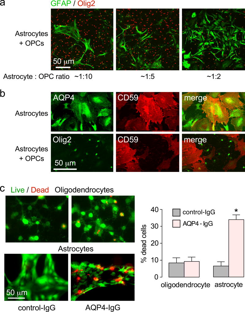
Characterization of rat oligodendrocyte-astrocyte cultures. a. GFAP and Olig2 immunofluorescence of oligodendrocyte-astrocyte cocultures at different cell ratios. Cells were cultured in the presence of 150 ng T3 for 3 days to induce maturation of oligodendrocyte precursor cells. b. AQP4 and CD59 immunofluorescence of astrocyte-oligodendrocyte cocultures. c. Complement-dependent cytotoxicity in pure oligodendrocyte and pure astrocyte cultures, measured by live/dead assay (calcein-AM/ethidium homodimer-1), following 2 h incubation with 30 µg/ml AQP4-IgG and 2% human complement. Percentage of dead cells summarized at the right (mean ± S.E.M., n=4, * P < 0.01).

