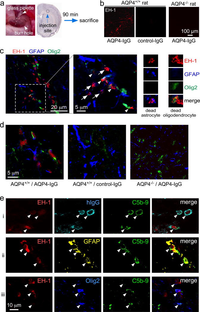
Figure 5.
Evidence for complement bystander mechanism for AQP4-IgG-induced oligodendrocyte injury in rat brain. a. AQP4-IgG (15 µg) (or control human IgG), human complement (26%) and fixable dead cell dye ethidium homodimer-1 (EH-1) (6 µM) in a 6-µl volume was injected in cortex and striatum of rat brain and rat were sacrificed at 90 min. b. Low-magnification micrographs showing dead cells (red EH-1 fluorescence) for studies done in AQP4+/+ and AQP4−/− rats. c. High-magnification confocal images of AQP4+/+ rat brain 90 min after injection of AQP4-IgG, complement and EH-1, showing dead astrocytes (white arrows) and nearby injured oligodendrocytes (white arrowheads). Expanded images on the right show dead (EH-1 positive) astrocyte and oligodendrocyte. d. Images as in panel c (center) showing representative fields from brains of AQP4+/+ and AQP4−/− rats following AQP4-IgG or control IgG injection. e. High-magnification confocal micrographs of brain from AQP4-IgG-injected AQP4+/+ rat showing colocalization (white arrowheads) of EH-1, hIgG and C5b-9 (row i), EH-1, GFAP and C5b-9 (row ii), and EH-1, Olig2 and C5b-9 (row iii).

