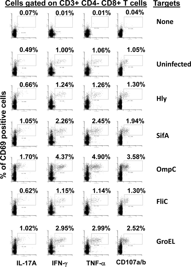
Fig 5. CD8+ T cell responses to S. Typhi proteins presented by targets infected with recombinant E. coli.

Ex vivo PBMC from a volunteer collected 42 days after immunization were co-cultured for 16–18 hrs. with autologous B-LCL targets infected at 1:30 MOI with one of the four recombinant E. coli expressing S. Typhi/Hly (Hly/SifA (SifA), Hly/FliC (FliC), Hly/GroEL (GroEL) and Hly/OmpC (OmpC)) or only Hly (control) proteins. After incubation, cells were stained and the ability of the PBMC to express one or more cytokines (IL-17A, IFN-γ and TNF-α) and/or CD107a/b molecules was evaluated by flow cytometry. Shown are the CD8+ T cell responses from a representative volunteer. Numbers represent the percentage of positive cells.
