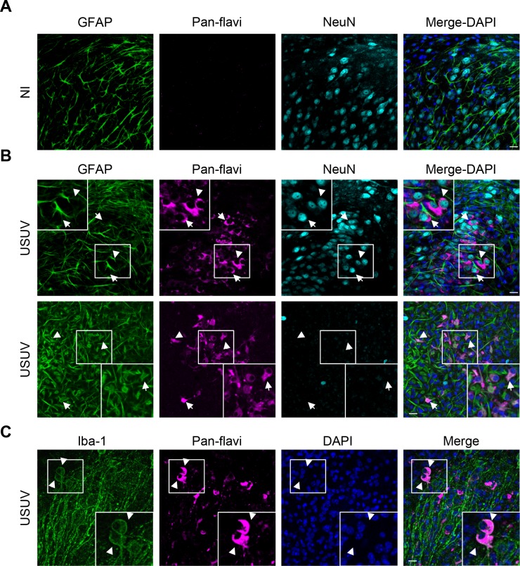
Fig 1. USUV infects efficiently organotypic murine brain slices.
Hippocampi slices obtained from 6 day old pups were infected with USUV (3×10⁵ infectious particles per slice). Five dpi, slices were fixed and subjected for indirect immunofluorescence using various neural cellular markers such as GFAP (astrocytes), NeuN (neurons) and Iba1 (microglia). (A) Non-infected (NI) slices did not show labeling by the anti-pan-flavivirus antibody, in contrast to USUV-infected samples (in magenta) (B and C). (B and C) White arrows show infected cells also expressing either NeuN (in cyan), GFAP or Iba1 (in green), indicating that USUV can infect and replicate in multiple types of neural cells in the murine brain. Nuclei are labeled with DAPI (in blue). Zoomed in panels of white boxes show co-labelling. Scale bars 20 μm.

