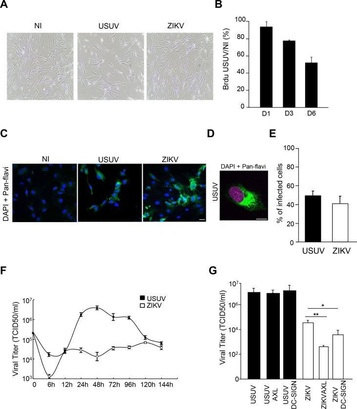
Fig 4. Comparative infections between USUV and ZIKV in primary human astrocytes.
Primary human astrocytes were infected with USUV or ZIKV at a MOI of 2. (A) Bright light images of control and infected astrocytes at 4 dpi show sparser cells in USUV-infected condition. (B) Cellular proliferation is affected by USUV infection. Astrocytes pre-treated with BrdU were infected with USUV at MOI 2 and assayed for BrdU at 1, 3 and 6 dpi. (C) Mock, USUV- and ZIKV-infected cells were fixed at 4 dpi and labeled with the pan-flavivirus antibody (in green) by indirect immunofluorescence. Strong labeling was observed in cells. Nuclei are labeled with DAPI (in blue). (D) Typical flavivirus labeling (in green) is observed at high magnification. Nuclei are labeled with DAPI (false colored in magenta). (E) Quantification of USUV and ZIKV-infected cells. (F) Kinetics of viral production in USUV- or ZIKV-infected astrocytes show difference in terms of replication and persistence between the two viruses. Supernatants from infected cells (MOI of 2) were collected at various time points and subjected to TCID50 measurement on Vero cells. (G) AXL and DC-SIGN do not mediate USUV internalization in human astrocytes. Cells were pre-incubated with anti-Axl or Anti-DC-SIGN prior to infection with USUV or ZIKV at MOI of 2. 4 dpi, supernatants were collected and viral replication assayed. Blocking antibodies only decreased ZIKV replication. (*p<0.05, **p<0.01). Scale bar 10 μm.

