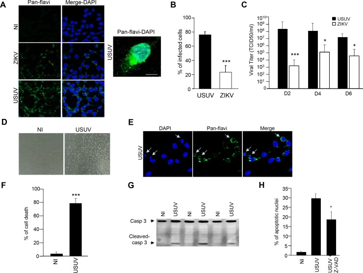
Fig 6. Effect of USUV infection on NSC survival.
(A) IPSc-derived NSCs were infected with USUV and ZIKV at a MOI of 2 and fixed at 2 dpi. Cells were labeled with the pan-flavivirus antibody (in green) by indirect immunofluorescence. Nuclei are labeled with DAPI (blue). Scale bar 10 μm. (B) Quantification of the percentage of USUV- and ZIKV-infected cells. (C) Supernatants from USUV- and ZIKV infected NSCs (MOI 2) were collected at 2, 4 and 6 dpi and subjected to TCID50 measurement on Vero cells. (D) Bright light images of control and USUV-infected NSCs at 4 dpi show rounded up cells in USUV-treated condition. (E) USUV-infected NSCS at 4 dpi and labeled with the anti-flavivirus antibody and DAPI show condensed nuclei. (F) Trypan blue assay (supernatant + adherent cells) showed that cell viability is affected in USUV-infected NSCs at 4 dpi. (G) Immunobloting analyses of cell extracts from mock or USUV-infected cells at 4 dpi show the generation of cleaved caspase-3 fragment, indicative of apoptosis. (H) NSCs were infected with USUV at a MOI of 2 with or without Z-VAD. 4 dpi, cells were labeled with the anti-flavivirus antibody and DAPI and condensed nuclei quantified. (*p<0.05, ***p<0.001).

